

Why Choose a Spine Surgery Microscope?
Why Choose a Spine Surgery Microscope? The answer begins with what the surgeon can see. In a narrow operative field, magnification and focused illumination can help reveal fine tissue planes, small vessels, and delicate nerve structures. A Spine Surgery Microscope brings these details into view while allowing the surgeon to work with controlled instruments. Small differences matter.
Microsurgery pioneer Dr. Gazi Yasargil is often associated with describing the microscope as the surgeon’s “third eye.” The phrase captures its purpose, but not its limits. A microscope can improve visualization; it cannot replace sound judgment, careful planning, or surgical experience. The patient’s anatomy may vary, and the view can change as the procedure progresses.
This introduction explores why spine teams use microscopes, how magnification and lighting support precision, and what practical factors shape their choice. These include working distance, field of view, positioning, and team ergonomics. A clear view is valuable. Yet it is not a guarantee of better results for every patient or procedure. The technology deserves careful consideration, not automatic praise. Its benefits depend on appropriate use, surgeon training, and the clinical situation. That balance is easy to overlook.
What Is a Spine Surgery Microscope?
Why Choose a Spine Surgery Microscope?
What Is a Spine Surgery Microscope?
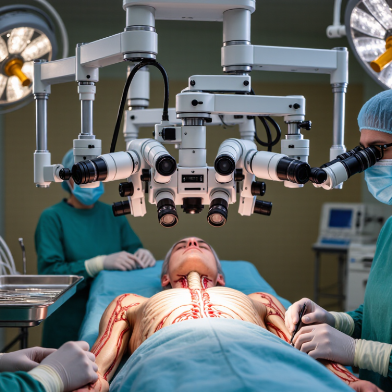
A spine surgery microscope is an operating-room instrument that magnifies the surgical field and directs bright light into it. Binocular eyepieces give the surgeon a detailed view of small structures, such as nerve roots, blood vessels, and disc fragments. That view can help guide precise work during procedures like microdiscectomy. It does not perform the surgery or replace the surgeon’s judgment. More detail is not automatically better; skill and careful planning still matter.
The SPORT study followed 1,244 people with lumbar disc herniation. At four years, its as-treated analyses found greater improvement among patients who had surgery than among those treated without surgery (Weinstein et al., Spine, 2008). The study compared treatment approaches, not microscope use, so it cannot show that a microscope alone improves results. That distinction matters when weighing claims about surgical tools.
Tips: Ask how the microscope supports your specific procedure, what alternatives are available, and how your surgeon will discuss risks and expected recovery. Get clear answers. Ask again if needed.
Why Choose a Spine Surgery Microscope? - What Is a Spine Surgery Microscope?
| Aspect | What It Means | Why It Matters in Spine Surgery | Important Consideration |
|---|---|---|---|
| What is a spine surgery microscope? | An operating microscope designed to provide magnified, illuminated views of the surgical field. | It can help the surgeon see small anatomical structures, including nerve roots and fine tissue planes, during selected procedures. | It supports visualization but does not replace surgical training, clinical judgment, or other imaging tools. |
| Magnification | Optical magnification makes the operative area appear larger to the surgeon. | A closer view can assist with precise work in a confined surgical corridor, such as during some decompression or microdiscectomy procedures. | The useful level of magnification depends on the task, field of view, and surgeon preference. |
| Illumination | Integrated lighting directs light into the operative field. | Focused illumination can improve visibility in deeper areas that may be difficult to light directly. | Lighting quality and positioning should be appropriate for the procedure and working distance. |
| Depth perception | Binocular optics provide a stereoscopic view when the microscope is properly set up. | A three-dimensional view can help the surgeon judge spatial relationships while working around delicate structures. | The benefit depends on correct adjustment, a clear line of sight, and the operator’s experience. |
| Typical applications | Microscope use may be considered for procedures involving fine dissection or a limited operative field. | Examples can include selected lumbar microdiscectomies, decompressions, and operations involving spinal tumors or intradural structures. | Use varies by procedure, surgeon, patient factors, and local practice; it is not required for every spine operation. |
| Ergonomics and positioning | The microscope can be positioned to provide a stable view while the surgeon works through the operative approach. | Appropriate positioning may help the surgeon maintain a comfortable viewing posture during fine work. | Setup and repositioning take time, and the microscope must not obstruct access or other equipment. |
| What it does not do | A microscope improves the surgeon’s optical view of the field; it does not independently identify anatomy or guide instruments. | Fluoroscopy, navigation, or other imaging may still be used when clinically indicated, particularly for procedures requiring instrument or implant localization. | The appropriate combination of visualization and imaging depends on the operation and the patient’s needs. |
| Choosing a microscope | Relevant factors include optical clarity, illumination, working distance, maneuverability, and compatibility with the operating room. | A suitable setup should support the procedures performed and allow the surgical team to work safely and efficiently. | Selection should account for the surgeon’s workflow, staff training, maintenance, and facility requirements. |

We have over 30 years of experience in designing complete hydraulic systems, repairs and distribution of fluid power equipment and parts.
FOLLOW US ON :
Contact Details
Address:
4020 SE 45th CT.
Ocala, FL 34480
Phone No:
Email:
sales@cunninghamfluidpower.com